Table of Contents
- What is Ultrasound?
- So, What Could Be Improved?
- Magnetomotive Ultrasound (MMUS) to the Rescue!
- What We’ve Accomplished So Far
- Publications
What is Ultrasound?
From emergency medicine to prenatal care, ultrasound is a highly versatile medical imaging modality capable of safe, cost-effective, and rapid diagnosis and treatment monitoring. A handheld transducer directs high-frequency (megahertz) sound waves into the human body. Reflections arise due to local changes in the speed of sound and/or density of tissue, which can be detected back at the probe. Much like sonar, the intensity and position of these reflections can be used to form an image.
So, What Could Be Improved?
Ultrasound works fantastically for many applications, but it does have a weakness: because images are formed by local changes in the sound speed and density of the tissue, ultrasound often cannot contrast objects with the same acoustic properties as the tissue surrounding them.
This is particularly relevant for thrombosis (blood clot) imaging. In this case, clinicians frequently have to infer the location of a clot rather than imaging it directly. Techniques include Doppler ultrasound which can visualize a narrowing of the blood flow through a vessel (a “stenosis”), or compression ultrasound wherein the clinician squishes the affected area and watches the recoil of the tissue. The “indirect” nature of these approaches means that ultrasound generally cannot provide quantitative information about the size or stiffness of a clot. This is a bummer because such information can be useful when determining how to treat clots, and how well a given course of treatment is working.

Magnetomotive Ultrasound (MMUS) to the Rescue!
Magnetomotive Ultrasound (MMUS) is a promising new technique in which the abilities of conventional ultrasound are enhanced by adding a “contrast agent.” More commonly used in other imaging modalities such as magnetic resonance imaging (MRI), contrast agents are molecules that “stick” to the part of the body the clinician wants to see, and then show up brightly in the image. In MMUS, I use magnetic nanoparticles as a contrast agent.
As shown in the diagram, two solenoid electromagnets are placed on either side of the ultrasound transducer to create a magnetic gradient force in the imaging area. I usually apply a sinusoidal force which causes any magnetic nanoparticles present in the imaging area to bounce up and down slightly. These nanoparticles are so small that the ultrasound system cannot image them directly. However, if the particles are attached to tissue, they cause the tissue to also bounce up and down. Although this motion is merely on the scale of tens of nanometers, it is detectable using phase-resolved image processing algorithms. Check out my Ph.D. advisor’s website for more information on magnetomotive imaging, and our magnetomotive ultrasound system in particular.
The long-term goal is to develop a method for labeling blood clots with magnetic nanoparticles, and then to image those clots with MMUS.
What We’ve Accomplished So Far

My students and I are currently working on developing MMUS for “elastometry” (stiffness sensing). I began by showing that nanoparticle displacement varies with the volume and stiffness of magnetically-labeled gelatin model blood clots embedded in a blood vessel mimic[2]. These results are important because the first step toward being able to measure the stiffness of a clot with MMUS is to demonstrate that MMUS is sensitive to changes in stiffness. The images displayed here show the same model blood clots two different ways: On the left, the clot is invisible in the conventional ultrasound image, but it shows up as a bright red region in the MMUS image on the right.
More recently, I conducted two studies toward the goal of actually measuring clot stiffnesses. In the first study, I created and validated a mathematical model to predict the motion of a single magnetic particle undergoing MMUS imaging[4]. Because the motion depends on the stiffness of the surrounding material, this is an important step toward elastometry. Then in the second study I demonstrated the use of a technique called “resonant acoustic spectroscopy” to directly measure the stiffnesses of gelatin clot-mimicking cylinders[5]. I swept the magnetic driving force over a range of frequencies, and used the resulting displacement data to determine the resonance frequency of the gelatin. This frequency depends on multiple factors including sample stiffness. By accounting for those other factors with a computational modeling approach called “finite element analysis” (FEA) I was able to use ultrasound measurements for quantitative elastometry.

In order for resonant acoustic spectroscopy to work, we need magnets capable of producing forces with both sufficient strength to actually wiggle the nanoparticles a detectable amount, and sufficiently high frequency to match all expected blood clot resonance frequencies. This will require a new, more nimble magnet design. Because finding the ideal solenoid configuration to meet our needs is a complicated optimization problem, my students and I saw another application for FEA. In a recent preprint[6], we presented and experimentally validated a simulation capable of predicting forces across a wide range of magnet geometries, driving frequencies, and more. We demonstrated that MMUS magnet performance could be enhanced through the strategic placement of permanent magnets, or by replacing two large solenoids with four smaller ones in specific orientations. These findings set out a roadmap for the next generation MMUS elastometry system.
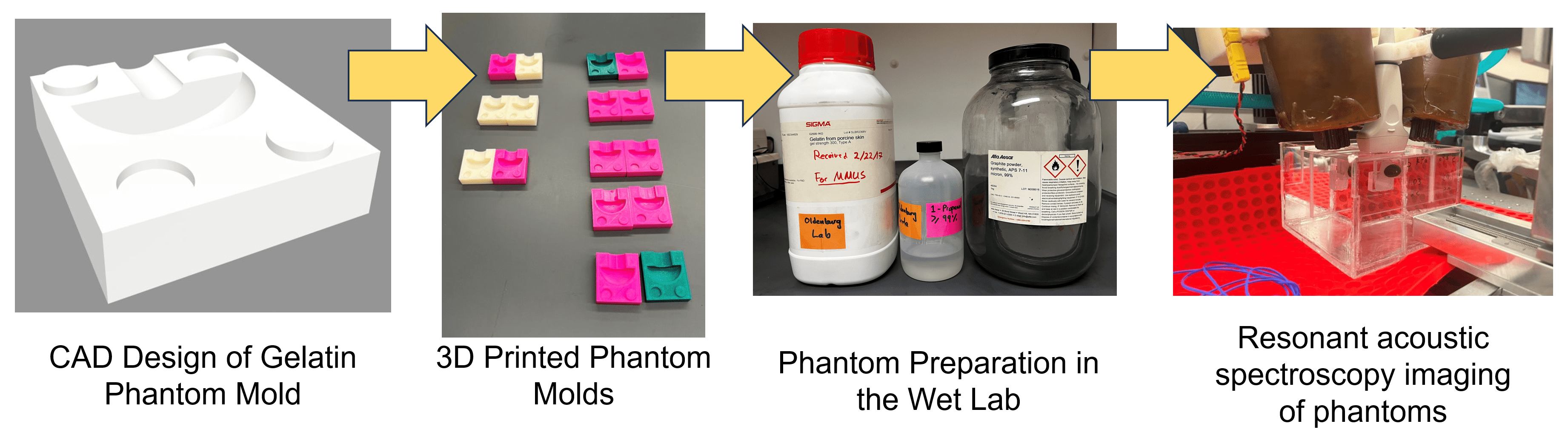
Furthermore, my students and I are extending the resonant acoustic spectroscopy approach to work for a more realistic range of gelatin clot-mimicking geometries. We created and validated a new model experimentally, and used it to predict the imaging hardware necessary for a range of future applications. Along the way we developed a faster method of creating phantoms.
More developments are in the works! If you want to take a deeper dive, feel free to peruse my publications:
Publications
*Indicates student co-author
[6] J. Nyakunu*, C. T. Piatnichouk*, H. C. Russell*, N. J. van Duijnhoven*, and B. E. Levy. “A Finite Element Analysis Model for Magnetomotive Ultrasound Elastometry Magnet Design with Experimental Validation,” under peer reivew, preprint arXiv:2408.07737 [physics.med-ph] (2024).
[5] B. E. Levy and A. L. Oldenburg, “Elastometry of Clot Phantoms via Magnetomotive Ultrasound-Based Resonant Acoustic Spectroscopy,” in Physics in Medicine & Biology, 67, 155010 (2022).
[4] B. E. Levy and A. L. Oldenburg, “Single Magnetic Particle Motion in Magnetomotive Ultrasound: An Analytical Model and Experimental Validation,” in IEEE Transactions on Ultrasonics, Ferroelectrics, and Frequency Control. doi: 10.1109/TUFFC.2021.3072867 (2021)
[3] D. Thapa, B. E. Levy, D. L. Marks and A. L. Oldenburg, “Inversion of Displacement Fields to Quantify the Magnetic Particle Distribution in Homogeneous Elastic Media from Magnetomotive Ultrasound,” in Physics in Medicine & Biology. doi: 10.1088/1361-6560/ab1f2b (2019)
[2] B. E. Levy, M. M. Hossain, J. M. Sierchio, D. Thapa, C. M. Gallippi and A. L. Oldenburg, “Effect of Model Thrombus Volume and Elastic Modulus on Magnetomotive Ultrasound Signal under Pulsatile Flow,” in IEEE Transactions on Ultrasonics, Ferroelectrics, and Frequency Control. doi: 10.1109/TUFFC.2018.2841774 (2018)
[1] M. M. Hossain, B. E. Levy, D. Thapa, A. L. Oldenburg and C. M. Gallippi, “Blind Source Separation Based Motion Detector for Imaging Super-Paramagnetic Iron Oxide (SPIO) Particles in Magnetomotive Ultrasound Imaging,” in IEEE Transactions on Medical Imaging. doi: 10.1109/TMI.2018.2848204 (2018)